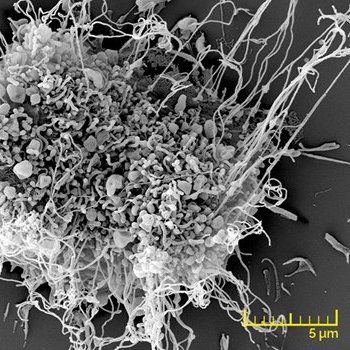
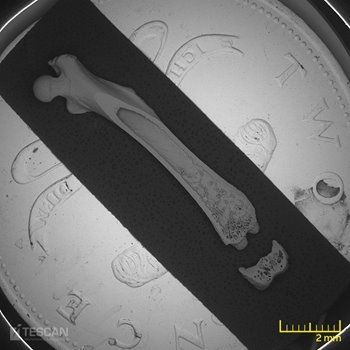
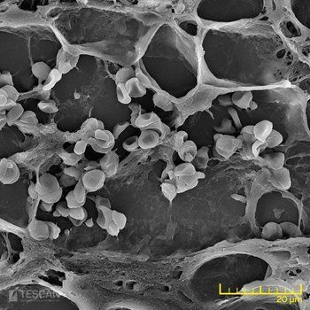
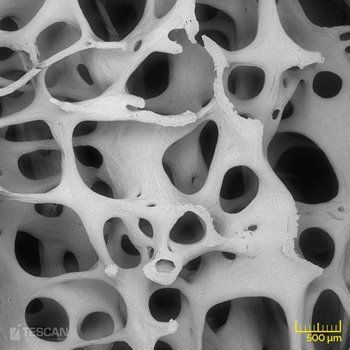
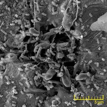
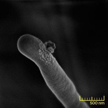
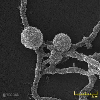
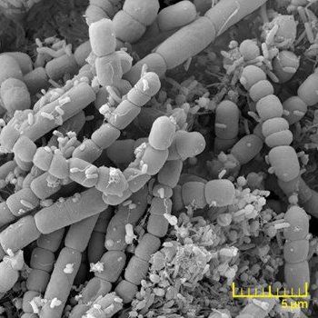
Scanning electron microscopy (SEM) has become an integral technology in biotechnology, life sciences and medical science research. Recent investigations of cell morphology, development of biocompatible materials, tissue engineering research, microbiology and many more heavily rely on advanced SEM imaging techniques. TESCAN develops and manufactures state-of-the-art electron microscopy solutions customised to every life science application. The broad range of dedicated instrumentation helps scientists and researchers in all fields to make stunning discoveries and move science forward.
Biomedical engineering
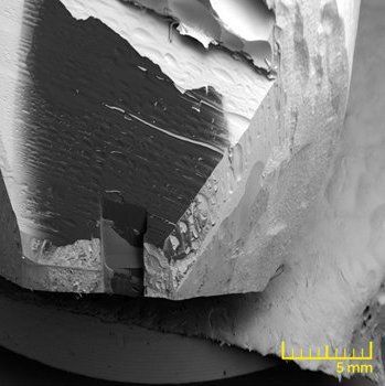
Biomedical engineering is one of the fastest growing research areas. It combines the latest concepts from medicine, biotechnology and engineering for designing a variety of technologies such as support matrices for cell growth, artificial tissue and implantable biomedical devices.
Slide title
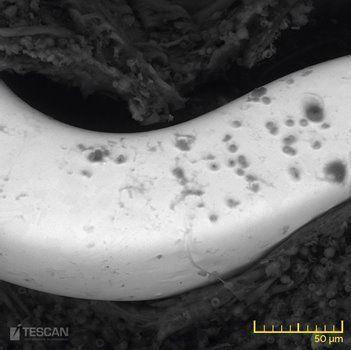
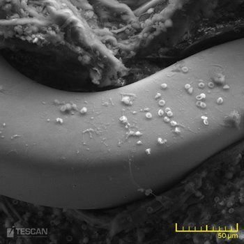
Contact layers under a solder ball imaged at 5 keV with the InBeam SE detector for topographic contrast (left), and the In-Beam f-BSE detector for material contrast (right).
Button
Slide title
Contact layers under a solder ball imaged at 5 keV with the InBeam SE detector for topographic contrast (left), and the In-Beam f-BSE detector for material contrast (right).
Button
Slide title
The beam deceleration improves imaging at low landing energies. Surface features in an Al flake become visible only at a landing energy of 500 eV (right). The multi-detector system enables simultaneous detection of BSE (left) and SE (right) signals.
Button
Slide title
The beam deceleration improves imaging at low landing energies. Surface features in an Al flake become visible only at a landing energy of 500 eV (right). The multi-detector system enables simultaneous detection of BSE (left) and SE (right) signals.
Button
Cell and Tissues Morphology
Cell membrane functions as a protection layer of the intracellular environment and together with cytoskeleton defines the outer shape of a cell.
Slide title
Contact layers under a solder ball imaged at 5 keV with the InBeam SE detector for topographic contrast (left), and the In-Beam f-BSE detector for material contrast (right).
Button
Slide title
Contact layers under a solder ball imaged at 5 keV with the InBeam SE detector for topographic contrast (left), and the In-Beam f-BSE detector for material contrast (right).
Button
Slide title
The beam deceleration improves imaging at low landing energies. Surface features in an Al flake become visible only at a landing energy of 500 eV (right). The multi-detector system enables simultaneous detection of BSE (left) and SE (right) signals.
Button
Slide title
The beam deceleration improves imaging at low landing energies. Surface features in an Al flake become visible only at a landing energy of 500 eV (right). The multi-detector system enables simultaneous detection of BSE (left) and SE (right) signals.
Button
Microbiology
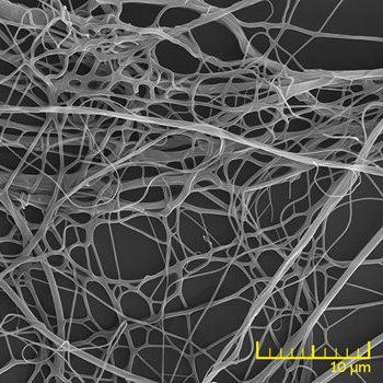
Microbial biofilms have been playing a major role in many processes such as infection, disease spreading and resistance to antibiotics.
Slide title
Contact layers under a solder ball imaged at 5 keV with the InBeam SE detector for topographic contrast (left), and the In-Beam f-BSE detector for material contrast (right).
Button
Slide title
Contact layers under a solder ball imaged at 5 keV with the InBeam SE detector for topographic contrast (left), and the In-Beam f-BSE detector for material contrast (right).
Button
Slide title
The beam deceleration improves imaging at low landing energies. Surface features in an Al flake become visible only at a landing energy of 500 eV (right). The multi-detector system enables simultaneous detection of BSE (left) and SE (right) signals.
Button
Slide title
The beam deceleration improves imaging at low landing energies. Surface features in an Al flake become visible only at a landing energy of 500 eV (right). The multi-detector system enables simultaneous detection of BSE (left) and SE (right) signals.
Button
Pharmaceuticals
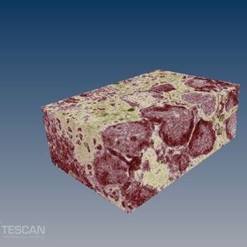
Electron microscopy plays an integral role in research and development of new drug formulations. Structure, particle size, porosity and presence of contaminants have been responsible for the final activity of the drug.
Slide title
Contact layers under a solder ball imaged at 5 keV with the InBeam SE detector for topographic contrast (left), and the In-Beam f-BSE detector for material contrast (right).
Button
Slide title
Contact layers under a solder ball imaged at 5 keV with the InBeam SE detector for topographic contrast (left), and the In-Beam f-BSE detector for material contrast (right).
Button
Slide title
The beam deceleration improves imaging at low landing energies. Surface features in an Al flake become visible only at a landing energy of 500 eV (right). The multi-detector system enables simultaneous detection of BSE (left) and SE (right) signals.
Button
Slide title
The beam deceleration improves imaging at low landing energies. Surface features in an Al flake become visible only at a landing energy of 500 eV (right). The multi-detector system enables simultaneous detection of BSE (left) and SE (right) signals.
Button
Plant and Animal Biology
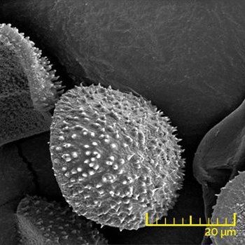
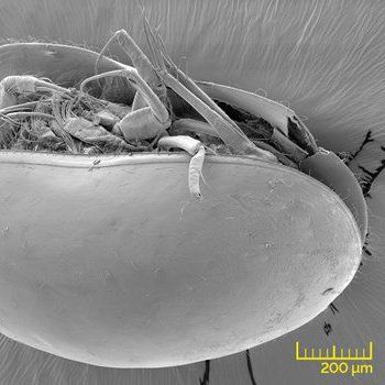
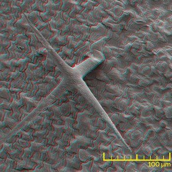
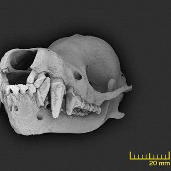
High resolution and large depth of focus makes SEM a great method for observing topography of biological samples, such as animal and plant fossils, bones, insects, plants and even small animals.
Slide title
Contact layers under a solder ball imaged at 5 keV with the InBeam SE detector for topographic contrast (left), and the In-Beam f-BSE detector for material contrast (right).
Button
Slide title
Contact layers under a solder ball imaged at 5 keV with the InBeam SE detector for topographic contrast (left), and the In-Beam f-BSE detector for material contrast (right).
Button
Slide title
The beam deceleration improves imaging at low landing energies. Surface features in an Al flake become visible only at a landing energy of 500 eV (right). The multi-detector system enables simultaneous detection of BSE (left) and SE (right) signals.
Button
Slide title
The beam deceleration improves imaging at low landing energies. Surface features in an Al flake become visible only at a landing energy of 500 eV (right). The multi-detector system enables simultaneous detection of BSE (left) and SE (right) signals.
Button
Slide title
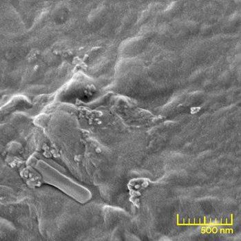
Image of an anode surface doped with Pt nanoparticles.
Button
Subcellular analysis
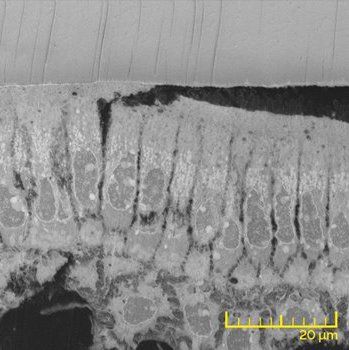
Subcellular analysis provides information hidden under the surface of cells and tissue. Typically this area of research has been a domain of transmission electron microscopy. However, SEM technology is becoming more popular in this field, due to emerging techniques available with scanning electron microscopy solutions.
Slide title
Contact layers under a solder ball imaged at 5 keV with the InBeam SE detector for topographic contrast (left), and the In-Beam f-BSE detector for material contrast (right).
Button
Slide title
Contact layers under a solder ball imaged at 5 keV with the InBeam SE detector for topographic contrast (left), and the In-Beam f-BSE detector for material contrast (right).
Button
Slide title
The beam deceleration improves imaging at low landing energies. Surface features in an Al flake become visible only at a landing energy of 500 eV (right). The multi-detector system enables simultaneous detection of BSE (left) and SE (right) signals.
Button
Environmental and Food Sciences
Recently, environmental science has become one of the fastest growing life science disciplines.
Slide title
Contact layers under a solder ball imaged at 5 keV with the InBeam SE detector for topographic contrast (left), and the In-Beam f-BSE detector for material contrast (right).
Button
Slide title
Contact layers under a solder ball imaged at 5 keV with the InBeam SE detector for topographic contrast (left), and the In-Beam f-BSE detector for material contrast (right).
Button
Slide title
The beam deceleration improves imaging at low landing energies. Surface features in an Al flake become visible only at a landing energy of 500 eV (right). The multi-detector system enables simultaneous detection of BSE (left) and SE (right) signals.
Button
Slide title
The beam deceleration improves imaging at low landing energies. Surface features in an Al flake become visible only at a landing energy of 500 eV (right). The multi-detector system enables simultaneous detection of BSE (left) and SE (right) signals.
Button
Need more information?
Subscribe to our newsletter
Thanks for contacting us. We will respond as soon as possible
There was an error sending your message. Try it later