Oficinas +34 91 148 8267
Tel. +34 682 715 975 (click para llamar)
Business Park Europa Empresarial
C/Rozabella 8,
Edificio Paris, oficina 12
28250 Las Rozas (Madrid)
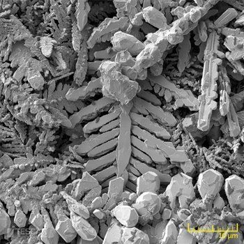
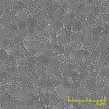
Contact layers under a solder ball imaged at 5 keV with the InBeam SE detector for topographic contrast (left), and the In-Beam f-BSE detector for material contrast (right).
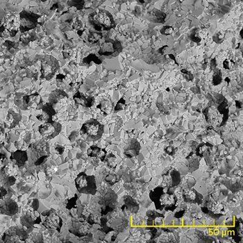
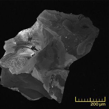
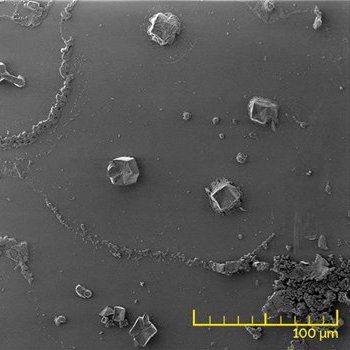
The beam deceleration improves imaging at low landing energies. Surface features in an Al flake become visible only at a landing energy of 500 eV (right). The multi-detector system enables simultaneous detection of BSE (left) and SE (right) signals.
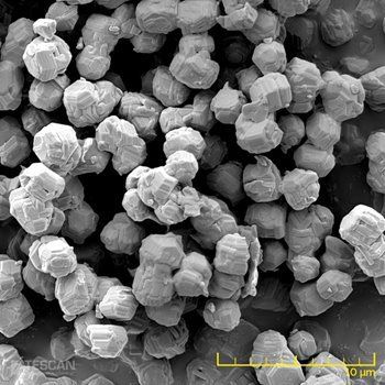
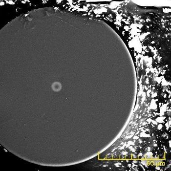
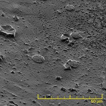
Image of an anode surface doped with Pt nanoparticles.
Contact layers under a solder ball imaged at 5 keV with the InBeam SE detector for topographic contrast (left), and the In-Beam f-BSE detector for material contrast (right).
The beam deceleration improves imaging at low landing energies. Surface features in an Al flake become visible only at a landing energy of 500 eV (right). The multi-detector system enables simultaneous detection of BSE (left) and SE (right) signals.
Image of an anode surface doped with Pt nanoparticles.
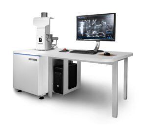
SEM analítico para aplicaciones rutinarias de caracterización de materiales, investigación y control de calidad a escala micrométrica.
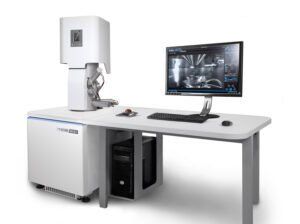
SEM analítico de alta resolución para aplicaciones rutinarias de caracterización de materiales, investigación y control de calidad a escala submicrónica.
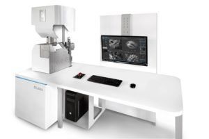
Crio-SEM UHR versátil para la caracterización de sus muestras biológicas y otras muestras sensibles al haz
Imágenes avanzadas de UHR SEM y STEM para la caracterización de sus muestras biológicas y sensibles al haz
FIB-SEM nanoanalítico versátil para ampliar sus capacidades de investigación de materiales.
Banco de trabajo de nanofabricación avanzada para su laboratorio de investigación.
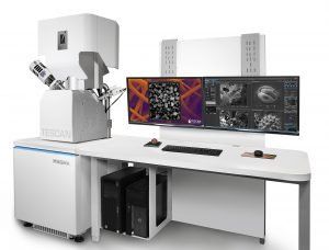
Una combinación única de Plasma FIB y UHR FE-SEM sin campo para la caracterización de materiales multi-escala.
Un micro-CT de resolución múltiple optimizado para un alto rendimiento, diversos tipos de muestras y flexibilidad para su investigación
TESCAN RISE combina la microscopía con focal Raman con la microscopía SEM (RISE) en un sistema de microscopio integrado.
Dirección:
Business Park Europa Empresarial
C/Rozabella 8,
Edificio Paris, oficina 12
28250 Las Rozas (Madrid)
Gracias por su interés
Envíenos su respuesta al cuestionario a socialmedia@zeppelinmet.com
Se ha producido un error al enviar su mensaje.
Inténtelo más tarde
Utilizamos cookies para asegurarnos de que le proporcionamos la mejor experiencia en nuestro sitio web. Consulte la página de privacidad para obtener más información.